Locking Compression Plate for Distal Tibia Fractures

Article Preview

Abstract:

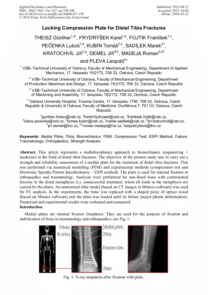
This article represents a multidisciplinary approach to biomechanics (engineering + medicine) in the field of distal tibia fractures. The objective of the present study was to carry out a strength and reliability assessment of a medial plate for the treatment of distal tibia fractures. This was performed via numerical modelling (FEM) and experimental methods (compression test and Electronic Speckle Pattern Interferometry – ESPI method). The plate is used for internal fixation in orthopaedics and traumatology. Analyses were performed for non-fused bone with comminuted fracture in the distal metaphysis (i.e. unsuccessful treatment, where all loads in the metaphysis are carried by the plate). An anatomical tibia model (based on CT images in Mimics software) was used for FE analysis. In the experiments, the bone was replaced with a shaped piece of spruce wood (based on Mimics software) and the plate was loaded until its failure (major plastic deformation). Numerical and experimental results were evaluated and compared.

You might also be interested in these eBooks

Info:

Periodical:

Pages:

359-366

Citation:

Online since:

February 2016

Export:

Price:

Permissions CCC:

Permissions PLS:

Сopyright:

© 2016 Trans Tech Publications Ltd. All Rights Reserved

Share:

Citation:

* - Corresponding Author

[1] B. Helgason, E. Perilli, E. Schileo et al., Mathematical relationships between bone density and mechanical properties, Clin. Biomech. 23 (2008) 135-146.

DOI: 10.1016/j.clinbiomech.2007.08.024

Google Scholar

[2] D.D. Cody, F.J. Hou, G.W. Divine et al., Short term in vivo precision of proximal femoral finite element modeling, Ann. Biomed. Eng. 28 (2000) 408-414.

DOI: 10.1114/1.1332085

Google Scholar

[3] J.H. Keyak. and Y. Falkinstein, Comparison of in situ and in vitro CT scan-based finite element model predictions of proximal femoral fracture load, Med. Eng. Phys. 25 (2003) 781-787.

DOI: 10.1016/s1350-4533(03)00081-x

Google Scholar

[4] F.E. Morgan, H. H. Bayraktar a M. T. Keaveny, Trabecular bone modulus–density relationships depend on anatomic site, J. Biomech. 36 (2003) 897-904.

DOI: 10.1016/s0021-9290(03)00071-x

Google Scholar

[5] M. Štamborská, M. Kvíčala, M. Losertová, Identification of the mechanical properties of high strength steel using digital image correlation, Adv. Mat. Res. 980 (2014) 122-126.

DOI: 10.4028/www.scientific.net/amr.980.122

Google Scholar

[6] K. Frydrýšek, F. Fojtík, O. Učeň et al., About the Verification of External Fixators Applied in Traumatology and Orthopaedics, Appl. Mech. Mat. 732 (2015) 153-160.

DOI: 10.4028/www.scientific.net/amm.732.153

Google Scholar

[7] K. Frydrýšek, J. Jořenek, O. Učeň et al., Design of External Fixators used in Traumatology and Orthopaedics – Treatment of Fractures of Pelvis and its Acetabulum, Procedia Eng. 48 (2012) 164-173.

DOI: 10.1016/j.proeng.2012.09.501

Google Scholar

[8] K. Frydrýšek, P. Koštial, K. Barabaszová et al., New Ways for Designing External Fixators Applied in Treatment of Open and Unstable Fractures, World Acad. Sci. Eng. Technol. 76 (2011) 697-702.

Google Scholar

[9] M. Vilimek and R. Sedláček. 2011. Methodology for Dynamic Testing of Plates for Proximal Tibia Osteosynthesis, Bull. Appl. Mech. 7 (2011) 12-14.

Google Scholar