p.325
p.329
p.333
p.337
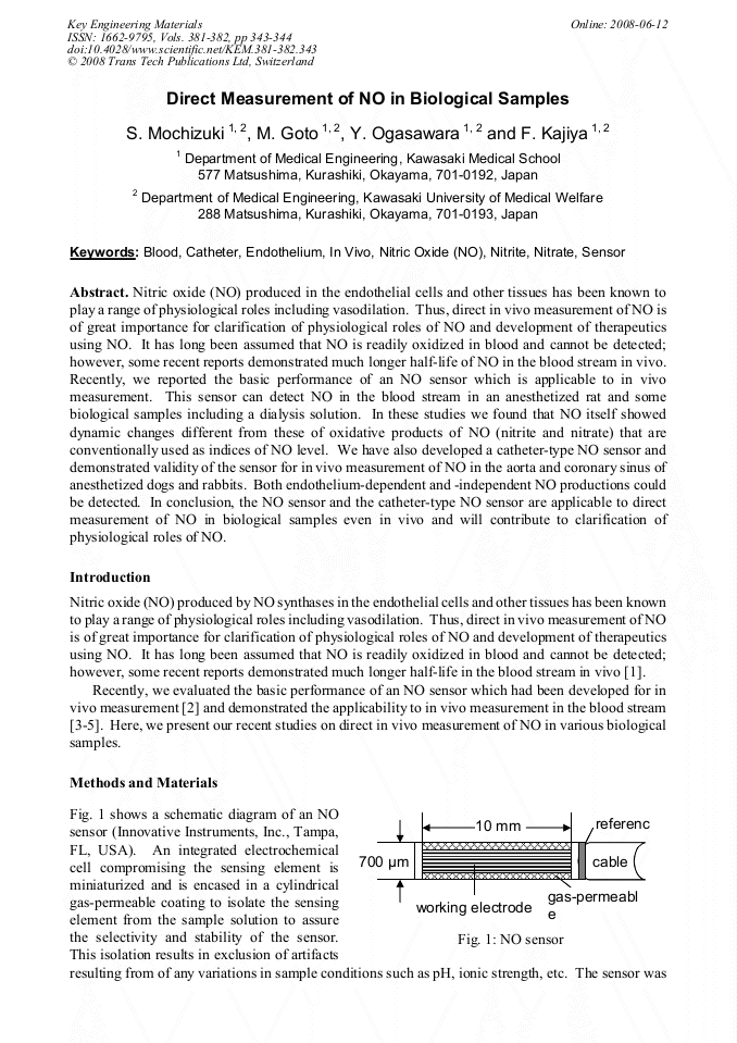
p.343
p.345
p.349
p.353
p.357
Direct Measurement of NO in Biological Samples

Abstract:
Nitric oxide (NO) produced in the endothelial cells and other tissues has been known to play a range of physiological roles including vasodilation. Thus, direct in vivo measurement of NO is of great importance for clarification of physiological roles of NO and development of therapeutics using NO. It has long been assumed that NO is readily oxidized in blood and cannot be detected; however, some recent reports demonstrated much longer half-life of NO in the blood stream in vivo. Recently, we reported the basic performance of an NO sensor which is applicable to in vivo measurement. This sensor can detect NO in the blood stream in an anesthetized rat and some biological samples including a dialysis solution. In these studies we found that NO itself showed dynamic changes different from these of oxidative products of NO (nitrite and nitrate) that are conventionally used as indices of NO level. We have also developed a catheter-type NO sensor and demonstrated validity of the sensor for in vivo measurement of NO in the aorta and coronary sinus of anesthetized dogs and rabbits. Both endothelium-dependent and -independent NO productions could be detected. In conclusion, the NO sensor and the catheter-type NO sensor are applicable to direct measurement of NO in biological samples even in vivo and will contribute to clarification of physiological roles of NO.
Info:
Periodical:
Pages:
343-344
Citation:
Online since:
June 2008
Authors:
Keywords:
Price:
Сopyright:
© 2008 Trans Tech Publications Ltd. All Rights Reserved
Share:
Citation: