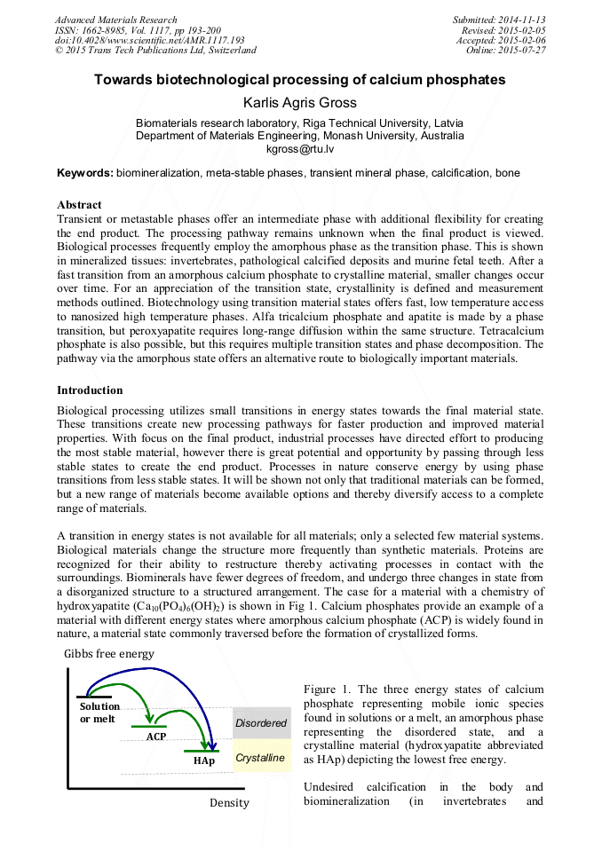
[1]
Zelenkova M, Sohnel O, Grases F. Ultrafine structure of the hydroxyapatite amorphous phase in noninfectious phosphate renal calculi. Urology 2012; 79: 968. e1-. e6.
DOI: 10.1016/j.urology.2011.11.020
Google Scholar
[2]
Gower LB, Amos FF, Khan SR. Mineralogical signatures of stone formation mechanisms. Urol Res 2010; 38: 281-92.
DOI: 10.1007/s00240-010-0288-z
Google Scholar
[3]
Hsu HHT, Camacho NP, Sun F, Tawfik S, Aono H. Isolation of calcifiable vesicles from aortas of rabbits fed with high cholesterol diets. Atherosclerosis 2000; 153: 337-48.
DOI: 10.1016/s0021-9150(00)00425-1
Google Scholar
[4]
Grynpas MD. Transient precursor strategy or very small biological apatite crystals? Bone 2007; 41: 162-64.
DOI: 10.1016/j.bone.2007.04.176
Google Scholar
[5]
Boskey AL. Amorphous calcium phosphate: The contention of bone. J Dental Res 1997; 76: 1433-36.
DOI: 10.1177/00220345970760080501
Google Scholar
[6]
Liu Y, Kim YK, Dai L, Li N, Khan SO, Pashley DH, et al. Hierarchical and non-hierarchical mineralisation of collagen. Biomaterials 2011; 32: 1291-300.
DOI: 10.1016/j.biomaterials.2010.10.018
Google Scholar
[7]
Lowenstam HA. Minerals formed by organisms. Science 1981; 211: 1126-31.
Google Scholar
[8]
Lowenstam HA. Phosphatic hard tissues of marine invertebrates: their nature and mechanical function, and some fossil implications. Chem Geol 1972; 9: 153-66.
DOI: 10.1016/0009-2541(72)90053-8
Google Scholar
[9]
Dorozhkin SV. Amorphous calcium orthophosphates: Nature, chemistry and miomedical applications. Int J Mater Chem 2012; 2: 19-46.
Google Scholar
[10]
Combes C, Rey C. Amorphous calcium phosphates: Synthesis, uses and properties in biomaterials. Acta Biomater 2010; 6: 3362-78.
DOI: 10.1016/j.actbio.2010.02.017
Google Scholar
[11]
Gross KA, Berndt CC, Herman H. Amorphous phase formation in plasma-sprayed hydroxyapatite coatings. J Biomed Mater Res 1998; 39: 407-14.
DOI: 10.1002/(sici)1097-4636(19980305)39:3<407::aid-jbm9>3.0.co;2-n
Google Scholar
[12]
Lopez EO, Mello A, Sendao H, Costa LT, Rossi AL, Ospina RO, et al. Growth of crystalline hydroxyapatite thin films at room temperature by tuning the energy of the RF-sputtering plasma. ACS Appl Mater Interfaces 2013; 5: 9435-45.
DOI: 10.1021/am4020007
Google Scholar
[13]
Gbureck U, Barralet JE, Radu L, Klinger HG, Thull R. Amorphous alpha-tricalcium phosphate: Preparation and aqueous setting reaction. J Am Ceram Soc 2004; 87: 1126-32.
DOI: 10.1111/j.1551-2916.2004.01126.x
Google Scholar
[14]
Miro S, Constantin JM, Bardeau JF, al. e. Raman spectroscopy study of damage induced in fluorapatite by swift heavy ion irradiation. . J Raman Spectrosc 2011; 42: 2036-41.
DOI: 10.1002/jrs.2955
Google Scholar
[15]
McElderry JDP, Zhu P, Mroue KH, Xu J, Pavan B, Fang M, et al. Crystallinity and compositional changes in carbonated apatites: Evidence from 31P solid-state, Raman and AFM analysis. J Solid State Chem 2013; 206: 192-98.
DOI: 10.1016/j.jssc.2013.08.011
Google Scholar
[16]
Yerramshetty JS, Akkus O. The associations between mineral crystallinity and the mechanical properties of human cortical bone. Bone 2008; 42: 476-82.
DOI: 10.1016/j.bone.2007.12.001
Google Scholar
[17]
Berg WA, Arnoldus CL, Teferra E, Bhargavan M. Biopsy of amorphous breast calcifications: Pathologic outcome and yield at stereotactic biopsy. Radiology 2001; 221: 495-503.
DOI: 10.1148/radiol.2212010164
Google Scholar
[18]
Gross KA, Andersons J, Misevicius M, Svirksts J. Traversing phase fields towards nanosized beta tricalcium phosphate. Key Engin Mater 2014; 587: 97-100.
DOI: 10.4028/www.scientific.net/kem.587.97
Google Scholar
[19]
Greene EF, Tauch S, Webb E, Amarasiriwardena D. Application of difuce reflectance infrared Fourier transform spectroscopy (DRIFTS) for the identification of potential diagenesis and crystallinity changes in teeth. Microchemical Journal 2004; 76: 141-49.
DOI: 10.1016/j.microc.2003.11.006
Google Scholar
[20]
Popovic L, de Waal D, Boeyens JCA. Correlation between Raman wavenumbers and P-O bond lengths in crystalline inorganic phosphates. J Raman Spectrosc 2005; 36: 2-11.
DOI: 10.1002/jrs.1253
Google Scholar
[21]
Brangule A, Gross KA. Effect of drying conditions on amorphous calcium phosphate. Key Engin Mater (2015).
Google Scholar
[22]
Gross KA, Komarovska L, Viksna A. Efficient zinc incorporation in hydroxyapatite through crystallization of an amorphous phase could extend the properties of zinc apatites. J Austral Ceram Soc 2013; 49: 129-35.
Google Scholar
[23]
Gross KA, Berndt CC, Herman H. Amorphous phase formation in plasma-sprayed hydroxyapaite coatings. J Biomed Mater Res 1998; 39: 407-14.
DOI: 10.1002/(sici)1097-4636(19980305)39:3<407::aid-jbm9>3.0.co;2-n
Google Scholar
[24]
Gross KA, Phillips MR. Identification and mapping of the amorphous phase in plasma-sprayed hydroxyapatite coatings using cathodoluminescnece microscopy. J Mater Sci: Mater Med 1998; 9: 797-802.
DOI: 10.1023/a:1008983809316
Google Scholar
[25]
Weaver JC, Wang Q, Miserez A, Tantuccio A, Stromberg R, Bozhilov KN, et al. Analysis of an ultra hard magnetic biomineral in chiton radular teeth. Materials Today 2010; 13: 42-52.
DOI: 10.1016/s1369-7021(10)70016-x
Google Scholar
[26]
Chatterji S, Wall JC, Fehhrey JW. Age-related changes in the orientation and particle size of the mineral phase in human femoral cortical bone. Calcifed Tissue International 1981; 33: 567-74.
DOI: 10.1007/bf02409493
Google Scholar
[27]
Paschalis EP, Betts F, DiCarlo E, Mendelsohn R, Boskey AL. FTIR microspectroscopic analysis of normal human cortical and trabecular bone. Calcif Tissue Int 1997; 61: 480-86.
DOI: 10.1007/s002239900371
Google Scholar
[28]
Saber Samandari S, Gross KA. Amorphous calcium phosphate offers improved crack resistance: A design feature from nature? Acta Biomater 2011; 7: 4235-41.
DOI: 10.1016/j.actbio.2011.06.048
Google Scholar
[29]
Le Geros RZ. Biodegradation and bioresorption of calcium phosphate ceramics. Clin Materials 1993; 14: 65-88.
Google Scholar
[30]
Tsuji T, Onuma K, Yamamoto A, Lijima M, Shiba K. Direct transformation from amorphous to crystalline calcium phosphate facilitated by motif-programmed artificial proteins. PNAS 2008; 105: 16866-70.
DOI: 10.1073/pnas.0804277105
Google Scholar
[31]
Liu Y, Kim YYK, Dai L, Li N, Khan SO, Pashley DH, et al. Hierarchical and non-hierarchical mineralisation of collagen. Biomaterials 2011; 2011: 1291-300.
DOI: 10.1016/j.biomaterials.2010.10.018
Google Scholar
[32]
Tiselius HG. The role of calcium phosphate in the development of Randall's plaques. Orolithiases 2013; 41: 369-77.
Google Scholar
[33]
Bertazzo S, Gentleman E, Cloyd KL, Chester AH, Yacoub MH, Stevens MM. Nano-analytical electron microscopy reveals fundamental insights into human cardiovascular tissue calcification. Nature Materials 2013; 12: 576-83.
DOI: 10.1038/nmat3627
Google Scholar
[34]
Heughebaert JC, Montel G. Revue de Physique Appliquee 1977; 12: 691-94.
Google Scholar
[35]
Root MJ. Inhibition of the amorphous calcium phosphate phase transformation reaction by polyphosphates and metal ions. Calcif Tissue Int 1990; 47: 112-16.
DOI: 10.1007/bf02555994
Google Scholar
[36]
Ajibola VO, Thomas SA. Transformation of amorphous calcium phosphate hydroxyapatite in the presence of some ions. Bull Chem Soc Ethiop 1997; 11: 19-24.
DOI: 10.4314/bcse.v11i1.21009
Google Scholar
[37]
LeGeros RZ. Formation and transformation of calcium phosphates: relevance to vascular calcification. Z Kardiol 2001; 90: 116-24.
Google Scholar
[38]
McCann TCA, Kearney RD, Buchheim W, Posner AS, Blumenthal NC. Amorphous calcium phosphate in casein micelles of bovine milk. Calcif Tissue Int 1983; 35: 821-23.
DOI: 10.1007/bf02405131
Google Scholar
[39]
Li Y, Wiliana T, Tam KC. Synthesis of amorphous calcium phosphate using various types of cyclodextrins. Mater Res Bull 2007; 42: 820-27.
DOI: 10.1016/j.materresbull.2006.08.027
Google Scholar
[40]
Kim CW, Yun YP, Lee HJ, Hwang YS, Kwon IK, Lee SC. In situ fabrication of alendronate-loaded calcium phosphate microspheres: controlled release for inhibitin of osteoclastogenesis. J Control Release 2010; 147: 45-53.
DOI: 10.1016/j.jconrel.2010.06.016
Google Scholar
[41]
Eanes ED. Thermochemical studies on amorphous calcium phosphate. Calc Tiss Res 1970; 5: 133-45.
DOI: 10.1007/bf02017543
Google Scholar
[42]
Vecbiskena L, Gross KA, Riekstina U, Thomas YCK. Formation of calcium-deficient hydroxyapatite via hydrolysis of nano-sized pure alpha-tricalcium phosphate. InterAcademia (2015).
DOI: 10.4028/www.scientific.net/amr.1117.201
Google Scholar
[43]
Raynaud S, Champion E, Bernache-Assollant D. Calcium phosphate apatites with variable Ca/P atmoic ratio II. Calcination and sintering. Biomaterials 2002; 23: 1073-80.
DOI: 10.1016/s0142-9612(01)00219-8
Google Scholar
[44]
Gross KA, Jersova A, Viksna A. Synthesis of peroxyapatite by hydrothermal processing. Key Engin Mater 2015; 631: 88-92.
DOI: 10.4028/www.scientific.net/kem.631.88
Google Scholar
[45]
Osite A, Gross KA, Viksna A, Poplausks R. Hydrothermally synthesized strontium peroxyapatite. InterAcademia (2015).
DOI: 10.4028/www.scientific.net/amr.1117.209
Google Scholar
[46]
Gross KA, Rozite E. Synthesis of tetracalcium phosphate at reduced temperatures. Key Engin Mater 2015; 631: 93-8.
DOI: 10.4028/www.scientific.net/kem.631.93
Google Scholar