Cranioplasty Based on Modern Methods for Personalized Design and Manufacturing

Article Preview

Abstract:

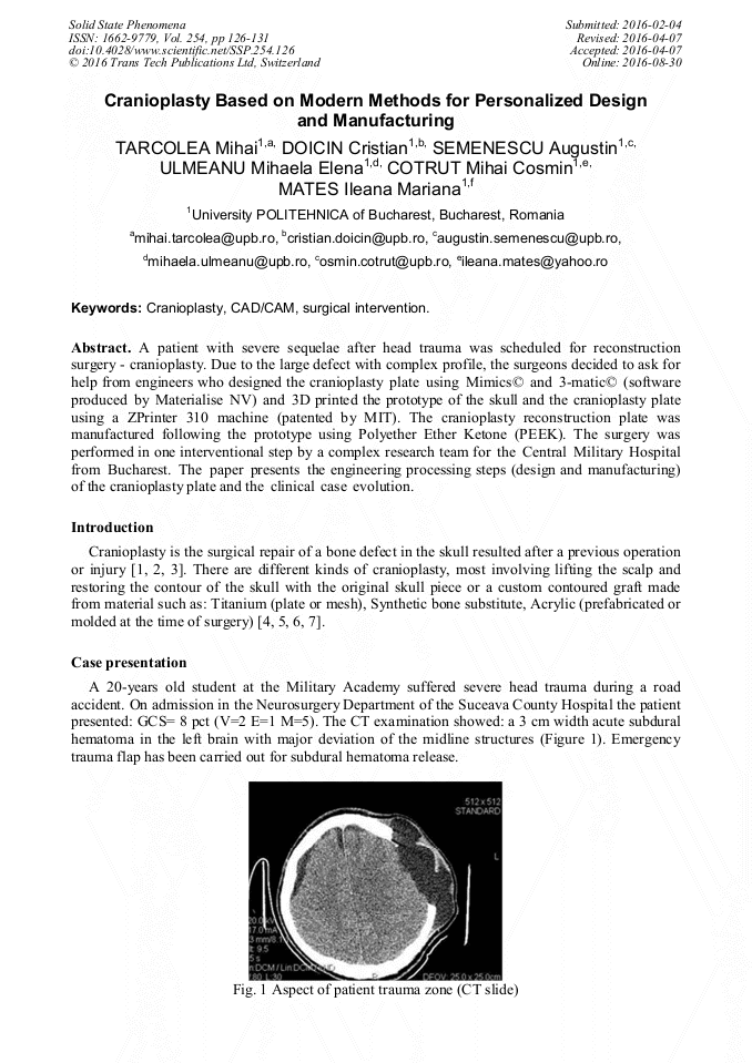
A patient with severe head trauma must was scheduled for cranioplasty. Due to the large defect with complex profile, the surgeons decided to ask for help from engineers who designed the cranioplasty plate using Mimics© and 3-matic© (software produced by Materialise NV) and 3D printed the prototype of the skull and the cranioplasty plate using a ZPrinter 310 machine (patented by MIT). The cranioplasty plate was manufactured following the prototype using Polyether Ether Ketone (PEEK). The surgery was a success as the first intervention using a complex research team for the Central Military Hospital from Bucharest. The paper presents the case evolution and the engineering processing steps (design and manufacturing) of the cranioplasty plate obtaining.

You might also be interested in these eBooks

Info:

Periodical:

Solid State Phenomena (Volume 254)

Pages:

126-131

Citation:

Online since:

August 2016

Export:

Price:

Permissions CCC:

Permissions PLS:

Сopyright:

© 2016 Trans Tech Publications Ltd. All Rights Reserved

Share:

Citation:

* - Corresponding Author

[1] C. Noufal Basheer et al., Cranioplasty following decompressive craniotomy in traumatic brain injury: Experience at Level - I apex trauma centre, Indian Journal of Neurotrauma, (2010).

DOI: 10.1016/s0973-0508(10)80029-2

Google Scholar

[2] Anastasia Tasiou et al., Cranioplasty optimal timing in cases of decompressive craniotomy after severe head injury: a systematic literature review, Interdisciplinary Neurosurgery, (2014).

DOI: 10.1016/j.inat.2014.06.005

Google Scholar

[3] K. Yoshida et al., Dynamics of cerebral blood flow and metabolism in patients with cranioplasty as evaluated by 133Xe CT and 31P magnetic resonance spectroscopy. Sponsored., J Neurol Neurosurg Psychiatry, (1996).

DOI: 10.1136/jnnp.61.2.166

Google Scholar

[4] Elisa Battistella et al., Cuttlefish bone scaffold for tissue engineering: a novel hydrothermal transformation, chemical-physical, and biological characterization., J Appl Biomater Funct Mater, (2012).

Google Scholar

[5] Gabriela Ciapetti et al., Enhancing Osteoconduction of PLLA-Based Nanocomposite Scaffolds for Bone Regeneration Using Different Biomimetic Signals to MSCs. Sponsored, Int J Mol Sci, (2012).

DOI: 10.3390/ijms13022439

Google Scholar

[6] S. Nakashima et al., In situ tissue engineering of the bile duct using polypropylene mesh- collagen tubes., Int J Artif Organs, (2007).

DOI: 10.1177/039139880703000110

Google Scholar

[7] Ng ZY, Nawaz I., Computer-designed PEEK implants: a peek into the future of cranioplasty? J Craniofac Surg., (2014).

DOI: 10.1097/scs.0b013e3182a2f7b6

Google Scholar

[8] Daniel R. Klinger et al., Autologous and Acrylic Cranioplasty: A Review of 10 Years and 258 Cases, World Neurosurgery, (2014).

Google Scholar

[9] Jayachandran Vekatesan et al., Chitosan Composites for bone tissue engineering – an overview. Sponsored. Mar Drugs, (2010).

Google Scholar