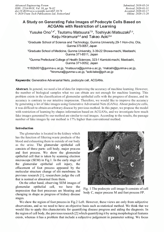
A Study on Generating Fake Images of Podocyte Cells Based on Acgans with Restriction of Learning

Article Preview

Abstract:

In general, we need a lot of data for improving the accuracy of machine learning. However, the number of biological samples what we can obtain are not enough for machine learning. This problem exists in the classification of glomerular epithelial cells with the progress of disease, and its accuracy is contrary to our intuitive impression. Therefore, we would like to improve the accuracy by generating a lot of fake images using Generative Adversarial Nets (GANs). About podocyte cells, it was difficult to obtain an arbitrary disease by previous method. In this paper, we propose the model with restriction of learning by shapes information based on ACGANs, and we investigate how much fake images generated by our method are similar to real images. According to the results, the passage number of fake images by our method is 17% higher than conventional method.

You might also be interested in these eBooks

Info:

Periodical:

Pages:

76-82

Citation:

Online since:

November 2020

Export:

Price:

Permissions CCC:

Permissions PLS:

Сopyright:

© 2020 Trans Tech Publications Ltd. All Rights Reserved

Share:

Citation:

* - Corresponding Author

[1] K. Asanuma, Molecular biology of podocyte, Juntendo Medical, 53 (2007) 11-19.

Google Scholar

[2] S. Motokawa, T. Matsuura, T. Aoki, Extract Cell Body Region from Microscopic Image of Podocyte and a Trial of Discrimination of Cellular Degeneration by Signal Regulatory Protein α and Diabetes Mellitus, Med Imag Tech, 33, 5 (2015), 208-216.

Google Scholar

[3] Y. Ono, T. Matsuura, T. Matsuzaki, et al., Developing and applying 2-step learning for analysis of glomerular epithelial cell images, JMEIS, 1, 1(2018), 7-15.

Google Scholar

[4] I. J. Goodfellow, J. Pouget-Abadie, M. Mirza, et al., Generative Adversarial Networks, CoRR, abs/ 1406.2661v1 (2014). URL https://arxiv.org/abs/1406.2661v1.

Google Scholar

[5] S. Takahashi, M. Tomioka, K. Hiromura, et al., SIRPα signaling regulates podocyte structure and function, Am J Physiol Rental Physiol, 305 (2013), 861-870.

Google Scholar

[6] S. Motokawa, T. Matsuura, T. Matsuzaki, et al., A Method for Automation of Extracting Foot Process Region from Podocyte Image, 3rd International Symposium of Gunma University Medical Innovation and 8th International Conference on Advanced Micro-Device Engineering, Kiryu, Japan, Dec. (2016).

Google Scholar

[7] A. Radford, L. Metz, S. Chintala, Unsupervised Representation Learning with Deep Convolutional Generative Adversarial Networks, CoRR, abs/ 1511.06434 (2015). URL https://arxiv.org/abs/1511.06434.

Google Scholar

[8] M. Mirza, S. Osindero, Conditional Generative Adversarial Nets, CoRR, abs/1411.1784 (2014). URL https://arxiv.org/abs/1411.1784.

Google Scholar

[9] A. Odena, C.olah, J.Shlens, Conditional Image Synthesis With Auxiliary Classifier GANs, CoRR, abs/1610.09585 (2016). URL https://arxiv.org/ abs/1610.09585.

Google Scholar

[10] Y. Ono, T. Matsuura, T. Matsuzaki, et al., Making fake images of podocyte for estimating the stages of kidney disease with machine learning, 5th International Symposium of Gunma University Medical Innovation and 9th International Conference on Advanced Micro-Device Engineering, Kiryu, Japan, Dec. (2016).

Google Scholar